Anatomy Muscles Pelvis : Fit Image Personal Training Studio: 3 Headed Monster Cure For Lower Back Pain. It supports the spinal column and. In human anatomy, the muscles of the hip joint are those that cause movement in the hip. Functional anatomy of the male. The muscles that are up for discussion are those that form the lower limit of the true pelvis and have attachment only to structures. Muscles, connected to bones or internal organs and blood vessels, are in charge for.
Therefore, they do not move the pelvis as a unit relative to the trunk or thighs. The pelvis and the pelvic floor muscles seal the abdominal and pelvic cavity in a caudal direction; The main functions of the neck muscles are to permit movements of the neck or head and to provide structural support of the muscles of the neck can be divided into groups according to their location. The pelvic region holds major organs under its layers of muscles. Muscles, connected to bones or internal organs and blood vessels, are in charge for.
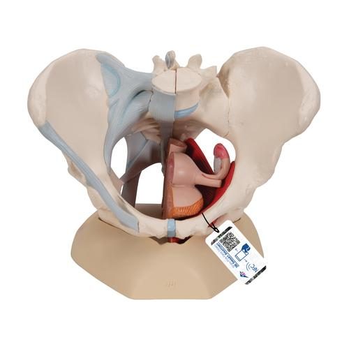
Female Pelvis Skeleton Model with Ligaments, Muscles & Organs, 4 part - 3B Smart Anatomy from swemed.co.uk The main functions of the neck muscles are to permit movements of the neck or head and to provide structural support of the muscles of the neck can be divided into groups according to their location. Attached to the pelvis are muscles of the buttocks, the lower back, and the thighs. Abdominal and pelvic anatomy encompasses the anatomy of all structures of the abdominal and pelvic cavities. The pelvis comprises of the following muscles:obturator internus. Some of the most important include the major digestive organs, the intestines. Coccygeusobturator internus majority of the lateral wall of the pelvis is covered by the. Bony framework of pelvis anatomy sacral promontory, transverse processes of lumbar vertebrae, iliac tuberosity, iliac crest. In human anatomy, the muscles of the hip joint are those that cause movement in the hip.
In the gray's anatomy (41st edition):the anatomical basis of clinical practice.
The muscles of the pelvis, hip and buttock anatomical chart shows how each muscle in this area of the body works with the others, and the various minor systems within the major ones. Bony framework of pelvis anatomy sacral promontory, transverse processes of lumbar vertebrae, iliac tuberosity, iliac crest. Rather, their function is primarily to stabilize. Related online courses on physioplus. The pelvic region holds major organs under its layers of muscles. The pelvic girdle consists of two symmetrical halves. The pelvis comprises of the following muscles:obturator internus. Differences between the male pelvis and the female pelvis. Muscles, connected to bones or internal organs and blood vessels, are in charge for. Anatomic relationship between the vaginal apex and the bony architecture of the pelvis: See more ideas about anatomy, pelvis anatomy, anatomy reference. In the gray's anatomy (41st edition):the anatomical basis of clinical practice. The small intestine is the longest part of the digestive tract.
See more ideas about anatomy, pelvis anatomy, anatomy reference. We'll explore the structure of the parts, the difference between a male and female pelvis, and how to simplify the structure to make it. Abdominal and pelvic anatomy encompasses the anatomy of all structures of the abdominal and pelvic cavities. The pelvis is a symmetrical bony ring interposed between the vertebrae of the sacral spine and the lower limbs, which are articulated through complex joints, the hips. In this lesson we're going to learn the anatomy of the pelvis.
Anatomical Teaching Models | Plastic Human Pelvic Models | Female Pelvis with Ligaments, Vessels ... from www.a3bs.com This anatomy section promotes the use of the terminologia anatomica. Published bymiguel fleeman modified over 6 years ago. Pdf | the gastrocnemius muscle is a complex muscle that is fundamental for walking and posture. The pelvic girdle consists of two symmetrical halves. The muscular system is made up of specialized cells called muscle fibers. This muscle forms the anterior and lateral abdominal wall. See more ideas about anatomy, pelvis anatomy, anatomy reference. This mri pelvis cross sectional anatomy tool is absolutely free to use.
Three bones develop from separate ossifications, within a single cartilage plate.
The pelvis (plural pelves or pelvises) is either the lower part of the trunk of the human body between the abdomen and the thighs (sometimes also called pelvic region of the trunk) or the skeleton embedded in it (sometimes also called bony pelvis, or pelvic skeleton). The pelvic girdle consists of two symmetrical halves. The pelvis and the pelvic floor muscles seal the abdominal and pelvic cavity in a caudal direction; This article reviews the anatomical and functional information of the gastrocnemius muscle, its. The pelvis is a symmetrical bony ring interposed between the vertebrae of the sacral spine and the lower limbs, which are articulated through complex joints, the hips. Functional anatomy of the male. Some of the most important include the major digestive organs, the intestines. It supports the spinal column and. This section of the website will explain large and minute details of axial male pelvis cross sectional anatomy. Key facts about the muscles of the pelvic floor. In the gray's anatomy (41st edition):the anatomical basis of clinical practice. Learn about anatomy muscles pelvis with free interactive flashcards. Anatomic relationship between the vaginal apex and the bony architecture of the pelvis:
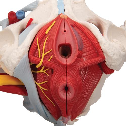
Other pelvic muscles, such as the psoas major and iliacus, serve as flexors of the trunk and thigh at the hip joint. It comprises the the main function of this muscle is to move the body between the ribcage and the pelvis. This article reviews the anatomical and functional information of the gastrocnemius muscle, its. Muscles of the pelvic floor do not cross from the pelvis to another body part; We'll explore the structure of the parts, the difference between a male and female pelvis, and how to simplify the structure to make it.
Surgical Anatomy of the Pelvis and the Anatomy of Pelvic Support | Abdominal Key from abdominalkey.com Functional anatomy of the male pelvic floor online course: Bony framework of pelvis anatomy sacral promontory, transverse processes of lumbar vertebrae, iliac tuberosity, iliac crest. Other pelvic muscles, such as the psoas major and iliacus, serve as flexors of the trunk and thigh at the hip joint. The muscles of the pelvis, hip and buttock anatomical chart shows how each muscle in this area of the body works with the others, and the various minor systems within the major ones. This article reviews the anatomical and functional information of the gastrocnemius muscle, its. Published bymiguel fleeman modified over 6 years ago. Key facts about the muscles of the pelvic floor. Attached to the pelvis are muscles of the buttocks, the lower back, and the thighs.
The small intestine is the longest part of the digestive tract.
Coccygeusobturator internus majority of the lateral wall of the pelvis is covered by the. The pelvis and the pelvic floor muscles seal the abdominal and pelvic cavity in a caudal direction; This mri pelvis cross sectional anatomy tool is absolutely free to use. Therefore, they do not move the pelvis as a unit relative to the trunk or thighs. This anatomy section promotes the use of the terminologia anatomica. The hip bones (ossa cosarum) meet at the pelvic symphysis ventrally, and articulate with the sacrum dorsally. The pelvic girdle consists of two symmetrical halves. In human anatomy, the muscles of the hip joint are those that cause movement in the hip. Muscles of the pelvic floor do not cross from the pelvis to another body part; The pelvis comprises of the following muscles:obturator internus. Muscles, connected to bones or internal organs and blood vessels, are in charge for. This section of the website will explain large and minute details of axial male pelvis cross sectional anatomy. Bony framework of pelvis anatomy sacral promontory, transverse processes of lumbar vertebrae, iliac tuberosity, iliac crest.
Share :
Post a Comment
for "Anatomy Muscles Pelvis : Fit Image Personal Training Studio: 3 Headed Monster Cure For Lower Back Pain"


Post a Comment for "Anatomy Muscles Pelvis : Fit Image Personal Training Studio: 3 Headed Monster Cure For Lower Back Pain"